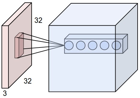
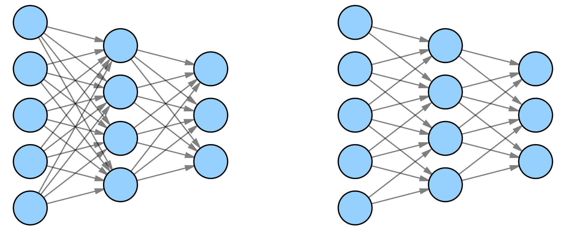
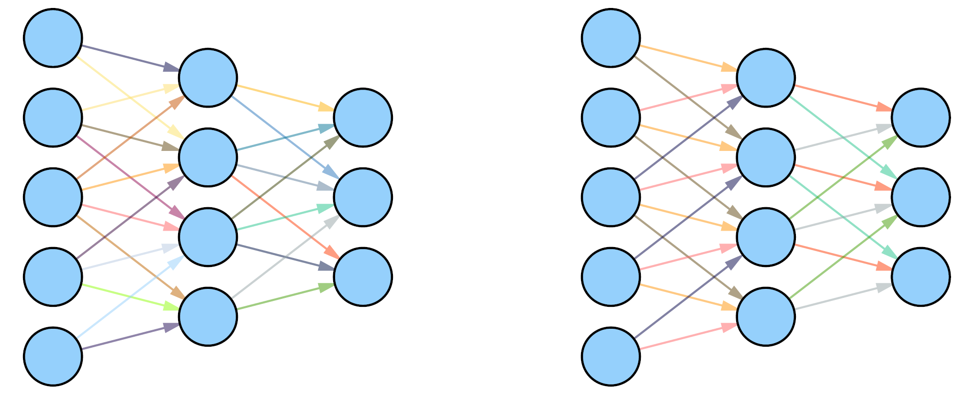
The other answer gives a good overview of the differences between MLPs and CNNs, and it includes 2 diagrams that attempt to illustrate the main differences between MLPs and CNNs, i.e. sparse connectivity and weight sharing. However, these diagrams do not clarify what a neuron in a CNN could be. A better diagram, which illustrates what a neuron is in a CNN, from a CNN and MLP perspective, is the following (taken from the famous article on CNNs).

Here, there are 2 main blocks (aka volumes): the orange block on the left (the input) and the blue/cyan volume on the right (the feature maps, i.e. the outputs of the convolutional layer, i.e. after the application of the convolutions with different kernels).
The circles in the visible stack of the cyan block represent the neurons (or, more precisely, their activations or outputs). We only see $k=5$ neurons stacked: this corresponds to the application of $k=5$ different kernels (i.e. weights) to that specific subset of the input (aka receptive field), hence the sparse connectivity of CNNs. So, these neurons, in the same stack, are looking at the same small subset of the input, but with different weights (i.e. kernels). The neurons, which are not shown in this diagram, that are on the same (vertical) 2d plane (known as feature map) of the same neuron (e.g. the first that we see from left to right) in the cyan volume are the neurons that share the same weights, i.e. we use the same kernel to produce their outputs.
So, in this biological/neuroscientific view of the CNN, when you apply the convolution (or cross-correlation) operation with 1 specific filter (or kernel), you are computing the activation (not to be confused with the activation function, which is used to compute the activation!) i.e. the output of multiple neurons, all of them share the same weights. You stack all these activations on the same 2d plane (known as feature map) of the output volume: note that this operation is just the convolution operation! When you compute the convolution with another kernel, you are again computing the activation of other multiple neurons, which share another different weight matrix, and so on and so forth.
Some authors prefer to use the term convolutional networks, i.e. without the term neural, probably because of this issue, i.e. it's not clear, especially to newcomers, what a neuron would be in a CNN, so the neuroscientific/biological view of CNNs is not always clear, although it's important to emphasize that CNNs were inspired by the visual cortext, so this biological interpretation could (and should) be more widely known or less confusing/misunderstood.
Now, let's address your question more directly.
Aren't the filters the same, in the way that they convert an "image" to a new "image" based on the weights that are in that filter? And that the next layer uses this new "image"?
The filters in a CNN correspond to the weights of an MLP.
A neuron in a CNN can be viewed as performing exactly the same operation as a neuron in an MLP. The big differences between a CNN and an MLP (as explained also in the other answer) are
Weight sharing: Some neurons (not all!) in the same convolutional layer share the same weights. The convolution (or cross-correlation) is the operation that implements this partial forward pass with the same weights for different neurons.
Neurons in a CNN only look at a subset of the input and not all inputs (i.e. receptive field), which leads to some notion of sparse connectivity
A convolutional layer, in a CNN, is composed of neurons in a 3d dimensional volume (or, more precisely, their activations are organized in a 3d volume), rather than a 1-dimensional one, as in an MLP.
CNNs may use subsampling (aka pooling)


